Tyson Foodservice operates as a critical division within Tyson Foods Inc., using its extensive protein production capabilities to supply a diverse array of foodservice clients across multiple sectors. As one of the largest protein providers in the US, Tyson Foods produces approximately 20% of the nation’s beef, pork, and chicken, which forms the foundation of
A hybrid convolutional neural network model for dental age estimation using buccal alveolar …
- Research
- Open access
- Published:
- Seyed Matin Mazloom Nezhad1,
- Erma Rahayu Mohd Faizal Abdullah1,
- Norliza Ibrahim2,
- Heba H. Bakhsh3,
- Uzair Ishtiaq1,4,
- Rabiah Al Adawiyah Rahmat2,
- Sarah AlMugairin3 &
- …
- Sara M. ElKhateeb5,6
BMC Oral Health volume 25, Article number: 1331 (2025) Cite this article
Abstract
Background
Age estimation is an essential task in medical dentistry and forensic sciences. Dental Age Estimation (DAE) is one of the most common methods for age estimation. Teeth are commonly used for age estimation because the schedules of tooth development and eruption are barely affected by the environment, nutrition, and socio-economic factors. However, conventional DAE methods are manually performed by clinicians, exposing bias and error to the estimated age. Moreover, the potentials of buccal alveolar bone level in DAE are rarely investigated. The aim of this study is to assess the effectiveness of buccal alveolar bone level of mandibular posterior teeth in DAE using Artificial Intelligence (AI) for children.
Methods
A total of 421 Dental Panoramic Tomography (DPT) of children ranging from 5 to 15 years of age were used to train multiple UNet segmentation models. Segmented images of teeth were extracted and fed into a Localization Convolutional Neural Network (CNN) to train them for measuring buccal alveolar bone level. Moreover, the buccal alveolar bone level measurements were then fed to the machine learning regression models for DAE.
Result
The transfer learning based UNet with VGG16 as its backbone achieved the best performance with an IoU score of 0.66 and the best performing Localization CNN achieved Mean Squared Error (MSE) of 0.0009 and (:{R}^{2}) score of 0.8266. The Support Vector Machine (SVM) regression model achieved the best mean absolute error of 0.99 year.
Conclusion
The results revealed the potential of buccal alveolar bone level for dental age estimation in children. Best performing model achieved an acceptable Mean Absolute Error (MAE) and similar results to Demirjian and London Atlas methods performed by human experts, showing promising results.
Trial registration
Not applicable.
Peer Review reports
Background
Human identification is a core task in clinical dentistry, forensic sciences and orthodontic treatment planning [1, 2]. There are many cases where the identification of a deceased person is critical, such as, fire explosions, chemical putrefaction, or any other form of physical damage. Dental Age Estimation (DAE) is one of the most common procedures to profile an unknown deceased person or undocumented individuals [3,4,5]. Many studies have shown the reliability of using human dental growth as an estimator of chronologic age [5]. Moreover, teeth are a great tool for human identification since they can remain intact longer than any other physical features. DAE is widely used as a form of age estimation of living persons in the absence of official birth records [6, 7]. One of the most established DAE methods is the assessment of tooth development using Dental Panoramic Tomography (DPT) [3, 8]. Many manual DAE techniques using DPT have been developed over the years [9,10,11].
One of the main difficulties in the field of dental age estimation is the variation in dental development and maturity between different populations and ethnic groups. Moreover, regionally, and ethnically specific data is required to be used for reliable and accurate age estimation techniques [12, 13]. Traditional forms of DAE are time-consuming and expensive and require human experts to ensure accurate and valid estimations [6]. Additionally, it has been shown that there are slight differences in the timing, duration, and dental maturity between sexes [14, 15]. Dental maturity, in general, is more advanced in females than males, which suggests that any form of DAE may need to take sex into account for more accurate estimations [14].
Over the last few years, many efforts have been made to use Artificial Intelligence (AI) and machine learning to help automate the age estimation process. Moreover, automated dental age estimation is an area of intense research, and more research has been conducted in this domain over the last few years [14]. For instance, Tao et al. [16] developed multiple machine learning algorithms and compared their performance against Demirjian’s and Willem’s methods on a dataset of 1636 cases. The study concluded that AI based approaches are more accurate than existing forms of dental age estimation. In another study, Farhadian et al. [17] developed another DAE model using the pulp-to-tooth ratio. The proposed method used a neural network to predict dental age. The study was conducted on a dataset of 300 Cone Beam Computed Tomography (CBCT) scans and showed better performance for neural networks than regression-based models.
Fan et al. [18], developed a hybrid deep learning model for DAE and compared it to manual methods on a large dataset of 15,915 DPT images ranging from 16 to 50 years old. Their approach achieved a Mean Absolute Error (MAE) of 2.61 years. They also evaluated the model on an extra independent dataset consisting of 100 samples which produced a MAE of 3.28 for males and 3.79 for females [18]. Their proposed model achieved better accuracy than manual methods.
In another study, Jacometti et al. [19] did a meta-analysis on the London Atlas method, a dental development chart proposed by AlQahtani et al. [20]. The London Method can estimate dental age from 28 weeks up to 23.99 years of age. They found the London Atlas method to be a reliable method providing acceptable error and bias on various datasets.
Koh et al. [21] studied 284 Cone-Beam Computed Tomography images of Malay and Chinese patients aged 20 and above and measured the buccal Alveolar Bone Level (ABL) to investigate its potential in dental age estimation. The authors concluded that ABL is highly correlated with chronological age and could be suitable for age estimation [21]. However, due to variations in dental development and maturity between different populations and ethnic groups, additional research is needed for different ethnic groups and populations.
Currently, there is limited AI based research supporting the use of ABL in dental age estimation for both adults and children, showing a big research gap in this area. To fill in this research gap, we proposed the current study, the development of an AI based dental age estimation system using ABL of mandibular posterior teeth for Saudi Children aged 5 to 15 years. The dental age is estimated using dental DPT images in four main steps, including, image processing, tooth detection using a UNet, ABL measurement using a Convolutional Neural Network (CNN) followed by a machine learning regression model for age estimation and compared it with human expert manual estimations. The aim of this study was to assess the effectiveness of buccal alveolar bone level of mandibular posterior teeth in DAE using AI for children. Major contributions of this paper are:
-
Propose a novel hybrid technique that uses buccal alveolar bone level for dental age estimation for children.
-
Evaluate the proposed hybrid model on a diverse dataset that contains the dental images of both male and female Saudi population of age ranging from 5 to 15 years.
-
Compare the performance of the proposed hybrid dental age estimation model, including the extraction of distinguishing features from UNet, VGG16 + UNet and Localization CNNs, with the baseline techniques.
-
To the best of our knowledge, this study is the first of its kind to use buccal alveolar bone level with localization CNN for the task of dental age estimation for children.
Methods
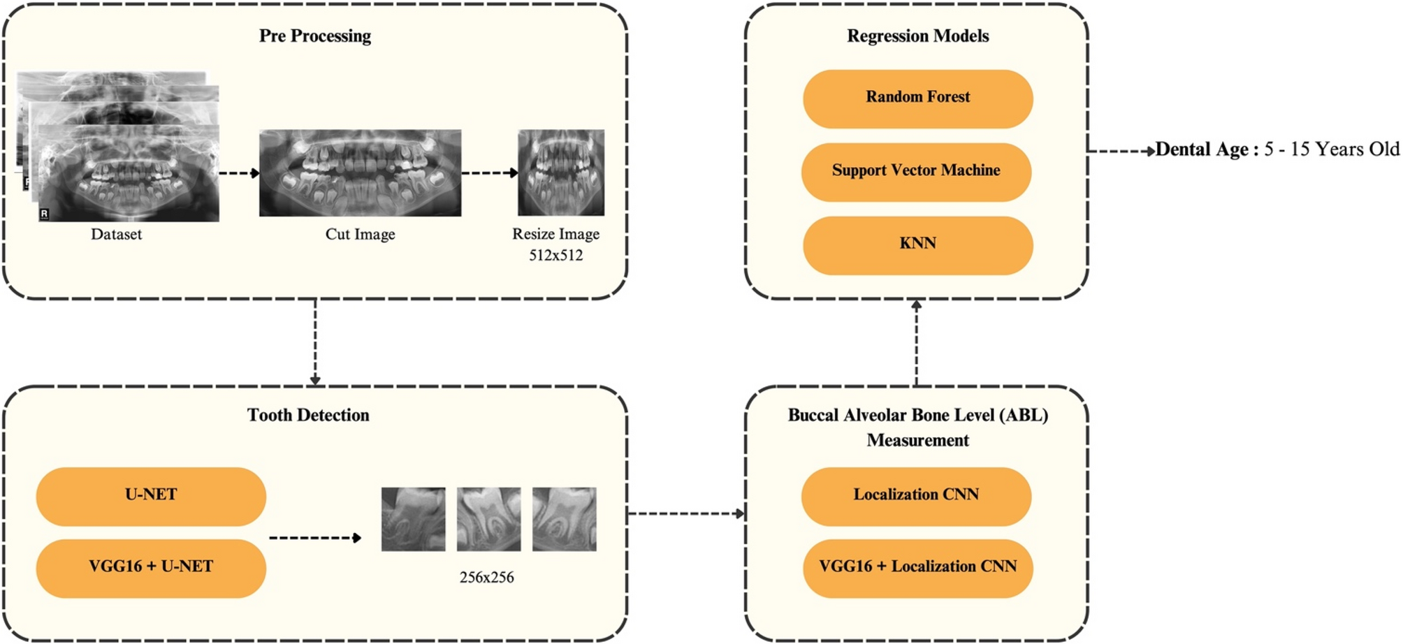
We proposed a multistage process to identify teeth, measure ABL and estimate dental age. The four main stages of our method are as follows: dataset preprocessing, tooth detection using deep learning models (UNet and VGG16 + UNet), ABL measurement using deep learning models (CNN and VGG16 + CNN) and dental age estimation using well known machine learning regression models. Lastly, all developed models were evaluated using multiple performance matrices and compared against 2 of the most commonly used manual DAE methods, the London Atlas and the Demirjian method. The developed process is illustrated in Fig. 1.

A multistage process to identify teeth, measure ABL and estimate dental age
Full size image
Dataset
In this study, a dataset of archived DPT from the faculty of dentistry, Princess Nourah bint Abdulrahman University (PNU) was obtained and used. The dataset contains 421 DPTs ranging from 5 to 15 years old, with 212 male and 209 female images. Table 1 provides the breakdown of the samples in the dataset. All images All images were anonymized and personally identifiable information on the DPT such as name and date of birth were removed and replaced with a random unique identifier number. Furthermore, manual age estimations using the London atlas and Demirjian method were done by a team of three dentists from PNU, trained and calibrated under the supervision of Dr. H.H.Bakhsh. The DPT samples were randomly divided among the dentists. The inter-rater reliability was 81% using Cohen’s kappa statistic (ρ-value < 0.001). A Google sheet form was used to record all the measurements.
Full size table
Preprocessing
In this stage, multiple operations were performed to prepare the dataset to be usable by the proposed models in subsequent stages. First, all images were converted into grayscale to reduce the number of dimensions in the dataset. This reduced the training time. Additionally, to focus on the most relevant regions within the images, the original images (2840 × 1532 pixels) were cropped around the region of interest, specifically, the teeth, reducing the image dimensions to 2638 × 1348 pixels. To further decrease computational load and enhance training efficiency, all cropped images were then resized to 512 × 512 pixels. This resolution was chosen as it significantly reduces image size while preserving sufficient detail for clear visualization of tooth boundaries, making it suitable for use with U-Net segmentation models.
Furthermore, data augmentation was performed to increase the number of images to help with the training process and reduce overfitting using translation [22]. Translation is a form of image augmentation that randomly shifts the image pixels on the X and Y axis by a bounded limit [23]. A bounded limit of 50 pixels was used on both the X and Y-axis. Furthermore, Image contrasts and brightness were adjusted to aid the image annotation process and help improve image clarity.
Teeth detection
In this stage, the main objective was to detect the tooth using a segmentation model, which was then used to cut the original larger images of 2840 × 1532 pixels down to smaller images of size 256 × 256 pixels that contain a single tooth at its center (Fig. 2). These cut images were then used to measure the ABL using a deep-learning model in stage 3. To achieve this, we trained and compared the performance of two multi-class segmentation models. A traditional UNet model and a transfer learning based model with VGG16 used as the feature extraction layer and multi-class UNet as output layer. UNet is a well-known model used for image segmentation with fast training times and it has been shown to work well with small datasets and to be effective with bio medical datasets [24,25,26]. For these reasons, the UNet model architecture was selected for this stage.

Cut images from tooth detection
Full size image
Due to the limited sample number, a second model was trained using the transfer learning technique. Transfer learning is a technique in which a pre-trained model is used for the feature extraction or input layer of the model [27]. This can reduce the training time and improve the model’s overall performance. For this purpose, a pre-trained VGG-16 model trained on the image net dataset, consisting of over 1 million images, was selected and used with UNet as the output layer. The two proposed models were then trained using the images from the previous stage. Both models were trained on images of size 512 × 512 pixels. The datasets were split into 70% training, 10% validation and 20% testing. Finally, the performance of these models was evaluated using different performance metrics, including, F1-Score (F1), accuracy (ACC), precision (PRE), specificity (SPE), sensitivity (SEN) and Intersection over Union (IoU). The mathematical formulation of the above metrics is as follows:
$$Accuracymathit=frac{mathit Nmathit umathit mmathit bmathit emathit rmathit;mathit omathit fmathit;mathit cmathit omathit rmathit rmathit emathit cmathit tmathit lmathit ymathit;mathit cmathit lmathit amathit smathit smathit imathit fmathit imathit emathit dmathit;mathit pmathit imathit xmathit emathit lmathit s}{mathit Tmathit omathit tmathit amathit lmathit;mathit nmathit umathit mmathit bmathit emathit rmathit;mathit omathit fmathit;mathit pmathit imathit xmathit emathit lmathit s}$$
(1)
$$Precision=frac{TP}{TP+FP}$$
(2)
$$F1=frac{2TP}{2TP+FP+FN}$$
(3)
$$Recall=frac{TP}{TP+FN}$$
(4)
$$IoU=frac{Area;of;overlap}{Area;of;union}$$
(5)
Where, TP = True Positive, FN = False Negative, TN = True Negative and FP = False Positive.
The generated segmentations from the models were then used to cut the original DPT images into multiple smaller 265 × 256 pixels images. To achieve this, the centroid of each label class was used to determine the center of the 256 × 256 images, placing the detected tooth at the center of the cut image. All generated images were then converted to grayscale using the same process detailed under the pre-processing section.
Buccal alveolar bone level measurement
At this stage, ABL was measured on the 256 × 256 pixels grayscale images. To achieve this, a localization CNN was used. As described by Koh et al. [21], the buccal alveolar bone level is the distance between Cemento-Enamel Junction (CEJ) and highest point of buccal bone as shown in Fig. 3. The model attempts to calculate the ABL for the first premolar, second premolar, first molar and second molar of the lower jaw if they are present and have been successfully detected and identified by the previous stage. First, the CNN model predicts the x and y coordinates of the CEJ and highest point of buccal bone for all the detected teeth on previous stage and then the ABL for each tooth is calculated by measuring the distance between these 2 points. It is important to note that in some DPT images specially for age ranges 5 to 10 years old may not contain some or all the above teeth due to the teeth not erupting yet. In such cases, the ABL of the available teeth were used at next stage for the training and estimation. Specially for 5 and 6 years old patients almost all image did not contain any of the teeth so no ABL meassurement could be done. In such cases the value of −1 was used in dataset to denote tooth not being erupted yet.

Buccal Alveolar Bone Level Measurement
Full size image
For the localization, a 22-layered CNN was used to estimate the CEJ and highest point of buccal bone points, as shown in Fig. 3, for the left side of each tooth. The CNN consisted of 10 layers with Leaky Rectified Linear Units (RELU) as activation functions, batch normalization, followed by a max pooling layer. The loss function, mean absolute error (MAE) was used as it focused more on smaller errors than mean squared error (MSE).
To assess the effectiveness of transfer learning in localization models as well as due to the limited sample number, a second model was trained using the transfer learning technique. For this purpose, a pre-trained VGG-16 model trained on the image net dataset, consisting of over 1 million images, was selected and used as feature selection layer of based model mentioned above.
The output layer returns 4 normalized coordinates for the 2 reference points CEJ and highest point of buccal bone on the left side of the tooth, each having an x and y coordinate. A total of 801 images were extracted from stage 1, annotated, augmented using translation as described in preprocessing and used to train the localization CNN. A data split of 70% training, 10% validation and 20% test was used. To assess the effectiveness of models they were evaluated using the following performance metrics: MAE, MSE and (:{R}^{2}).
$$MSE=frac{sum_{i=o}^{N-1}left|y_i-{widehat y}_iright|^2}N$$
(6)
$$MAE=frac{sum_{i=0}^{N-1}left|y_i-{widehat y}_iright|}N$$
(7)
$$R^2=1-frac{sumleft(y_i-{widehat y}_iright)^2}{sumleft(y_i-overline yright)^2}$$
(8)
Where, (:hat {{y}}_{i}) is predicted coordinates by model, (:{y}_{i}) is ground truth coordinates of each image and (:overline{y}) is mean of all (:{y}_{i}) values.
Dental age estimation
At this stage, the normalized ABL measurements of the first molar and premolar as well as second molar and premolar teeth measured in the previous stage are passed through various well-known machine learning regression models, including Support Vector Machine (SVM), Random Forest (RF), and K-Nearest Neighbors (K-NN). Each model was trained with two different variations of sex-neutral and sex-specific. After cleaning and removing any unusable datasets from the previous stages, a total of 196 data sets were used for training and testing. The process involved removing any datasets containing medical appliances, overlapping teeth preventing measurement of ABL and low quality or unclear images preventing ABL measurement. Also, to ensure the test results were accurately representing the models’ prediction quality, the test set was split in a balanced manner containing the same number of samples for each class or age. This is important since the model has significantly higher accuracy for some age ranges. For example, majority of images for 5 and 6 year olds does not contain any teeth thus making the prediction very simple and easy with very rare few cases containing 1 or 2 of the tooth erupted. Because of the reasons mentioned above the test split was done in a balanced manner.
Due to the smaller data points, the dataset was split into 70% training, 10% validation and 20% test set. To assess the performance of trained models, the following performance metrics were used: mean absolute error (MAE), mean square error (MSE) and root mean square error (RMSE) were used. To further evaluate the effectiveness of the models, they were then compared with 2 of the most popular manual DAE methods of London Atlas and Demirjian method.
$$MSE=frac{sum_{i=0}^{N-1}left|y_i-{widehat y}_iright|^2}N$$
(9)
$$MAE=frac{sum_{i=0}^{N-1}left|y_i-{widehat y}_iright|}N$$
(10)
$$RMSE=sqrt[2]{frac{sum_{i-0}^{N-1}left|y_i-{widehat y}_iright|^2}N}$$
(11)
Where, (:hat {{y}}_{i}) is predicted age by the model, (:{y}_{i}) is ground truth age.
Results
The detailed results and findings for each of the proposed models are presented in subsections below.
Tooth detection
The proposed segmentation models were implemented using Tensorflow. The trained UNet models were assessed using five different performance measurements: accuracy (ACC), precision, F1-Score (F1), recall and intersection over union (IoU).
The performance of segmentation models is presented in Table 2. The best performance was achieved using the transfer learning approach with UNet and VGG16 as its backbone, with an IoU score of 0.66, accuracy of 98.45%, F1 score of 0.98, recall of 0.98, and precision of 0.98. Followed by the traditional UNet model achieving an IoU score of 0.60, accuracy of 98.08%, F1 score of 0.98, recall of 0.98 and precision of 0.98. Both models are effective at segmenting teeth in DPT images for the purpose of tooth detection in this study, with the transfer learning approach showing superior performance.
Full size table
Based on the presented results, the transfer learning model outperformed the traditional UNet model.
Buccal alveolar bone level measurement
2 CNN models were trained for the task of point localization, A 22-layered CNN as well a transfer learning-based model using a pre-trained VGG16 model for its feature selection layer. To measure the performance of the trained model, 3 performance measures were used, MSE, MAE and coefficient of determination ((:{R}^{2})).
Table 3 presents the results achieved by proposed models. The best model achieved a MSE score of 0.0009, MAE of 0.0225 and a (:{R}^{2}) score 0.8266. The transfer learning model did not show any performance improvements compared to the base method with a MSE of 0.0009, MAE of 0.0235 and (:{R}^{2})of 0.8066.
Full size table
Dental age estimation regression models
In total, six regression models were trained using the SVM, RF and K-NN models, with each model having two variations consisting of sex-neutral and sex-specific models. To assess the performance of the trained models, four performance metrics were used: MAE, MSE and root mean square error (RMSE).
Where SN stands for sex-neutral, and SS stands for sex-specific.
Based on Table 4, it is observed that the sex-specific SVM model was the best performing model, achieving a MAE of 0.99, MSE of 1.86 and RMSE of 1.36, followed by the sex-neutral SVM model with a MAE of 0.99, MSE of 1.88 and RMSE of 1.37. In contrast, the lowest performing model, sex-neutral RF, reached a MAE of 1.33, a MSE of 2.22 and an RMSE of 1.49. Based on the results, it can also be seen that for each respective model, the sex-specific variant achieved slightly better results further supporting the differences in dental maturity between boys and girls.
Full size table
To further assess, the performance of the trained models, we compared the best performing trained model against 2 of the existing traditional methods of DAE the London Atlas Method and the Demirjian method as shown in Table 5. On our dataset the London Atlas method achieved the best results with a MAE of 0.79, MSE of 1.13 and RMSE of 1.06 with second best performing approach the Demirjian method with MAE of 0.91, MSE of 1.36 and RMSE of 1.16, followed closely by the sex-specific SVM model that achieved a MAE of 0.99, MSE of 1.86 and RMSE of 1.36.
Full size table
It can be seen from the results that the best performing model, achieved very close performance to existing methods and shows promising results as a potentially new alternative method to be used for dental age estimation in cases where other methods are not applicable or to be combined with existing methods.
Discussion
Dental age estimation is one of the most important forms of age estimation in forensics and dentistry, and can be a quick and effective way to estimate a person’s age in the absence of official documents such as in processing undocumented immigrants, determining age at time of death and ensuring eligibility of athletes to compete [5, 28]. Additionally, people of different ethnicities tend to have different tooth growth patterns, suggesting further research in various regional and ethnic groups is needed [29].
Previous studies have developed and assessed many manual and AI based methods for DAE. The aim of this study was to develop a new and novel method of DAE for children using buccal alveolar bone level of the mandibular teeth. To aid with the measurement and collection of ABL measurements used for DAE, a hybrid multistage system was developed. A tooth detection stage using a segmentation model to help identify the teeth followed by localization stage to help measure ABL. The ABL measurements were then fed into 3 regression models consisting of RF, SVM and K-NN. To further assess the impact of sex on dental age, 2 variations of each model was trained, a sex-natural and a sex-specific model.
Effectiveness of deep learning models is largely dependent on the quality and size of dataset being used. Transfer learning is a technique were a pretrained model is used to improve model performance and overcome small dataset size [27]. Recent studies have shown better performance when using transfer learning, with VGG16 showing significant improvement in performance on medical images [30]. Thus, for the tooth detection stage, 2 segmentation models were trained, a traditional UNet and a transfer learning based UNet with VGG16 as its backbone. The VGG16 UNet achieved the best performance with an IoU score of 0.66 followed by the traditional UNet model with an IoU score of 0.60 further supporting the effectiveness transfer learning in segmentation tasks.
At next stage, 2 localization CNNs were used to measure the ABL distance in pixels. A basic CNN as well as a transfer learning based CNN using VGG16 as its feature extractor to also assess effective of transfer learning in localization task. The base CNN model achieved the best results with a MSE of 0.0009, MAE of 0.0225 and (:{R}^{2}) score 0.8266. The transfer learning approach did not perform better than base model unlike the segmentation task, this could be due to the fact that the image net dataset does not contain images similar or related to the dataset used for the localization stage, thus not having any positive impact on training time or model performance.
Finally, the buccal alveolar bone level measurements of the lower jaw were used to train 6 machine learning models. The best performing model sex-specific SVM achieved a MAE of 0.99, MSE of 1.86 and RMSE of 1.36 followed closely by the sex-neutral SVM with a MAE of 0.99, MSE of 1.88 and RMSE of 1.37. When compared to manual traditional methods done by human experts it achieved comparable results using the same dataset. The London Atlas method had the best results with a MAE of 0.79, MSE of 1.13 and RMSE of 1.06 followed by the Demirjian method with MAE of 0.91, MSE of 1.36 and RMSE of 1.16. This matches with the findings of a recent pilot studies done on Indian population where London Atlas’s estimations were closer to chronological age than Demirjian method [31]. While the developed method achieved similar result as traditional method, it is able to perform DAE at a much faster rate, being able to make estimation in less than 1 s on modern hardware after training is done, while traditional methods take on average around 5 min to perform. Additionally, AI methods can be to run faster by increasing processing power or by increasing number of computers to processes multiple patients at once. Which is not possible with a manual method. Moreover, automated methods are less prune to human error and can provide more consistent results. Additionally, of all the trained models, the sex-specific variants achieved slightly better results than sex-neutral variants suggesting that for the most accurate age estimation in children, sex may need to be considered due to differences in dental maturity [14].
The findings of the present study show that buccal alveolar bone level measurements could be used as a predictor of dental age and could be used as an alternative method for dental age estimation in absence of other methods or be combined with existing methods for more accurate estimation as shown by Gelbrich et al. [32] when they achieved the highest precision from the average of the London Atlas and Willems’s method [32]. Additionally, alveolar bone level (ABL) may be useful for estimating age in cases where the mandible is fragmented or partially destroyed. However, this study has several limitations, including a small sample size and reliance on dental panoramic tomography (DPT). As a two-dimensional (2D) imaging modality, DPT is prone to geometric distortion and magnification errors, which can compromise measurement accuracy. Moreover, overlapping anatomical structures or the presence of medical appliances may obscure the view of the teeth, further limiting precise evaluation. In contrast, three-dimensional (3D) imaging techniques such as cone-beam computed tomography (CBCT) can overcome many of these limitations by providing more accurate and detailed anatomical representations. Finally, the age range of 5 to 15 years does not provide insights into the effectiveness of ABL for DAE in adults. Future research should aim to increase the sample size, include a broader age range encompassing adults, and utilize 3D imaging modalities for improved accuracy.
Conclusion
This study examined the effectiveness of buccal alveolar bone level for dental age estimation using a hybrid AI approach on DPT images. The developed method can estimate dental age on DPT for age ranges of 5 to 15 years old by measuring the ABL of the first and second premolar as well as first and second molar tooth of the lower jaw if the aforementioned teeth are present. The best performing model, sex-specific SVM, achieved an acceptable MAE and comparable results to Demirjian and London Atlas methods performed by human experts with the advantage of being automated, making estimations in less time and being able to perform multiple estimations at the same time. Application of ABL for DAE in children demonstrated promising results when mandibular teeth are available. The proposed method could be used in conjunction with other methods or when other methods are not applicable, and can be expanded upon in future research for adults.
Data availability
The datasets generated or analyzed during the current study are not publicly available to protect patient privacy but are available from the corresponding author on reasonable request.
Abbreviations
- DAE:
-
Dental Age Estimation
- AI:
-
Artificial Intelligence
- CBCT:
-
Cone Beam Computed Tomography
- CNN:
-
Convolutional Neural Network
- MAE:
-
Mean Absolute Error
- MSE:
-
Mean Squared Error
- RMSE:
-
Root Mean Square Error
- DPT:
-
Dental Panoramic Tomography
- SVM:
-
Support Vector Machine
- RELU:
-
Leaky Rectified Linear Units
- K-NN:
-
K-Nearest Neighbours
- RF:
-
Random Forest
- ABL:
-
Buccal Alveolar Bone Level
- PNU:
-
Princess Nourah bint Abdulrahman University
- CEJ:
-
Cemento-Enamel Junction
References
-
Kahm SH, Kim J-Y, Yoo S, Bae S-M, Kang J-E, Lee SH. Application of entire dental panorama image data in artificial intelligence model for age estimation. BMC Oral Health. 2023. https://doi.org/10.1186/s12903-023-03745-x.
Article PubMed PubMed Central Google Scholar
-
Bagherian A, Sadeghi M. Assessment of dental maturity of children aged 3.5 to 13.5 years using the Demirjian method in an Iranian population. J Oral Sci. 2011;53(1):37–42.
PubMed Google Scholar
-
Uzuner FD, Kaygısız E, Darendeliler N. Defining Dental Age for Chronological Age Determination. Post Mortem Examination and Autopsy – Current Issues From Death to Laboratory Analysis. 2018. https://doi.org/10.5772/intechopen.71699.
-
Willems G, Van Olmen A, Spiessens B, Carels C. Dental age estimation in Belgian children: demirjian’s technique revisited. J Forensic Sci. 2001;46(4):893–5.
PubMed Google Scholar
-
Lewis JM, Senn DR. Dental age estimation utilizing third molar development: a review of principles, methods, and population studies used in the United States. Forensic Sci Int. 2010;201(1–3):79–83.
PubMed Google Scholar
-
Milošević D, Vodanović M, Galić I, Subašić M. Automated Estimation of chronological age from panoramic dental X-ray images using deep learning. Expert Syst Appl. 2022;189. https://doi.org/10.1016/j.eswa.2021.116038.
-
Al-Emran S. Dental age assessment of 8.5 to 17 year-old Saudi children using demirjian’s method. J Contemp Dent Pract. 2008;9(3):64–71.
PubMed Google Scholar
-
Ozveren N, Serindere G. Comparison of the applicability of Demirjian and Willems methods for dental age estimation in children from the Thrace region, Turkey. Forensic Sci Int. 2018;285:38–43.
PubMed Google Scholar
-
Sehrawat JS, Singh M. Willems method of dental age estimation in children: a systematic review and meta-analysis. J Forensic Leg Med. 2017;52:122–9.
PubMed Google Scholar
-
Nayyar A, Babu B, Krishnaveni B, Devi M, Gayitri HC. Age estimation: current state and research challenges. J Med Sci. 2016. https://doi.org/10.4103/1011-4564.196348.
Article Google Scholar
-
Wang J, Fan L, Shen S, Sui M, Zhou J, Yuan X, et al. Comparative assessment of the Willems dental age estimation methods: a Chinese population-based radiographic study. BMC Oral Health. 2022. https://doi.org/10.1186/s12903-022-02418-5.
Article PubMed PubMed Central Google Scholar
-
Ranasinghe S, Perera J, Taylor JA, Tennakoon A, Pallewatte A, Jayasinghe R. Dental age estimation using radiographs: towards the best method for Sri Lankan children. Forensic Sci Int. 2019;298:64–70.
PubMed Google Scholar
-
Galibourg A, Cussat-Blanc S, Dumoncel J, Telmon N, Monsarrat P, Maret D. Comparison of different machine learning approaches to predict dental age using Demirjian’s staging approach. Int J Legal Med. 2021;135(2):665–75.
PubMed Google Scholar
-
Vila-Blanco N, Varas-Quintana P, Tomás I, Carreira MJ. A systematic overview of dental methods for age assessment in living individuals: from traditional to artificial intelligence-based approaches. Int J Legal Med. 2023;137(4):1117–46.
PubMed PubMed Central Google Scholar
-
Moorrees CFA, Fanning EA, Hunt EE. Age variation of formation stages for ten permanent teeth. J Dent Res. 1963;42(6):1490–502.
PubMed Google Scholar
-
Tao J, Wang J, Wang A, Xie Z, Wang Z, Wu S et al. Dental Age Estimation: A Machine Learning Perspective. The International Conference on Advanced Machine Learning Technologies and Applications (AMLTA2019). Advances in Intelligent Systems and Computing. 2020;722–33. https://doi.org/10.1007/978-3-030-14118-9_71.
-
Farhadian M, Salemi F, Saati S, Nafisi N. Dental age Estimation using the pulp-to-tooth ratio in canines by neural networks. Imaging Sci Dentistry. 2019;49(1). https://doi.org/10.5624/isd.2019.49.1.19.
-
Fan F, Ke W, Dai X, Shi L, Liu Y, Lin Y, et al. Semi-supervised automatic dental age and sex estimation using a hybrid transformer model. Int J Legal Med. 2023;137(3):721–31.
PubMed Google Scholar
-
Jacometti V, Sato CM, Meireles DA, Silva RHA. Age estimation using London atlas methodology: a systematic review and meta-analysis. Forensic Sci Int. 2023. https://doi.org/10.1016/j.forsciint.2022.111532.
Article PubMed Google Scholar
-
AlQahtani SJ, Hector MP, Liversidge HM. Brief communication: the London atlas of human tooth development and eruption. Am J Phys Anthropol. 2010;142(3):481–90.
PubMed Google Scholar
-
Koh KK, Tan JS, Nambiar P, Ibrahim N, Mutalik S, Khan Asif M. Age estimation from structural changes of teeth and buccal alveolar bone level. J Forensic Leg Med. 2017;48:15–21.
PubMed Google Scholar
-
Lin S-Y, Chang H-Y. Tooth numbering and condition recognition on dental panoramic radiograph images using CNNs. IEEE Access. 2021;9:166008–26.
Google Scholar
-
Xu M, Yoon S, Fuentes A, Park DS. A comprehensive survey of image augmentation techniques for deep learning. Pattern Recogn. 2023;137. https://doi.org/10.1016/j.patcog.2023.109347.
-
S UK, J P, S KSR. Systematic Review of Deep Learning Models for Dental Images. 2023 7th International Conference on Computing Methodologies and Communication (ICCMC) 2023;287–91. https://doi.org/10.1109/ICCMC56507.2023.10083729.
-
Seo H, Badiei Khuzani M, Vasudevan V, Huang C, Ren H, Xiao R et al. Machine learning techniques for biomedical image segmentation: an overview of technical aspects and introduction to state-of‐art applications. Med Phys. 2020;47(5). https://doi.org/10.1002/mp.13649.
-
Yüksel AE, Gültekin S, Simsar E, Özdemir ŞD, Gündoğar M, Tokgöz SB, et al. Dental enumeration and multiple treatment detection on panoramic X-rays using deep learning. Sci Rep. 2021. https://doi.org/10.1038/s41598-021-90386-1.
Article PubMed PubMed Central Google Scholar
-
Bal-Ghaoui M, El Yousfi Alaoui MH, Jilbab A, Bourouhou A. U-net transfer learning backbones for lesions segmentation in breast ultrasound images. International Journal of Electrical and Computer Engineering (IJECE). 2023. https://doi.org/10.11591/ijece.v13i5.pp5747-5754.
Article Google Scholar
-
Shen S, Liu Z, Wang J, Fan L, Ji F, Tao J. Machine learning assisted cameriere method for dental age estimation. BMC Oral Health. 2021. https://doi.org/10.1186/s12903-021-01996-0.
Article PubMed PubMed Central Google Scholar
-
Lin Y, Maimaitiyiming N, Sui M, Abuduxiku N, Tao J. Performance of the London atlas, Willems, and a new quick method for dental age estimation in Chinese Uyghur children. BMC Oral Health. 2022. https://doi.org/10.1186/s12903-022-02652-x.
Article PubMed PubMed Central Google Scholar
-
Albashish D, Al-Sayyed R, Abdullah A, Ryalat MH, Ahmad Almansour N. Deep CNN Model based on VGG16 for Breast Cancer Classification. 2021 International Conference on Information Technology (ICIT). 2021;805–10. https://doi.org/10.1109/icit52682.2021.9491631.
-
Chowdhry A, Kapoor P, Bhargava D, Bagga DK, Mehta A. Comparison of demirjian’s comprehensive chart with the London atlas of tooth development in children and adolescents: a pilot study. Forensic Sci Res. 2023;8(4):332–7.
PubMed PubMed Central Google Scholar
-
Gelbrich B, Carl C, Gelbrich G. Comparison of three methods to estimate dental age in children. Clin Oral Invest. 2019;24(7):2469–75.
Google Scholar
Download references
Acknowledgements
Not applicable.
Funding
This work was supported by Faculty of Dentistry, Universiti Malaya (UM.0000371/KWJ.SR) and Fundamental Research Grant Scheme (FRGS), Ministry of Higher Education, Malaysia (Grant: FP1212020/FRGS/1/2020/SKK0/UM/02/23).
Ethics declarations
Ethics approval and consent to participate
The research was authorized and exempted from the IRB review by the Institutional Review Board of Princess Nourah bin Abdulrahman University (HAP-01-R-059). The requirement for informed consent was waived due to the retrospective nature of the study and the use of non-human subject data (dental panoramic radiographs). However, general informed consent for the use of clinical data for research purposes was obtained from all patients, or from the guardians of patients under 16 years of age, at the time of treatment at the University. The research was conducted in accordance with the Declaration of Helsinki.
Consent for publication
Not applicable.
Competing interests
The authors declare no competing interests.
Additional information
Publisher’s Note
Springer Nature remains neutral with regard to jurisdictional claims in published maps and institutional affiliations.
Rights and permissions
Open Access This article is licensed under a Creative Commons Attribution-NonCommercial-NoDerivatives 4.0 International License, which permits any non-commercial use, sharing, distribution and reproduction in any medium or format, as long as you give appropriate credit to the original author(s) and the source, provide a link to the Creative Commons licence, and indicate if you modified the licensed material. You do not have permission under this licence to share adapted material derived from this article or parts of it. The images or other third party material in this article are included in the article’s Creative Commons licence, unless indicated otherwise in a credit line to the material. If material is not included in the article’s Creative Commons licence and your intended use is not permitted by statutory regulation or exceeds the permitted use, you will need to obtain permission directly from the copyright holder. To view a copy of this licence, visit http://creativecommons.org/licenses/by-nc-nd/4.0/.
Reprints and permissions
About this article

Cite this article
Mazloom Nezhad, S.M., Mohd Faizal Abdullah, E.R., Ibrahim, N. et al. A hybrid convolutional neural network model for dental age estimation using buccal alveolar bone level for Saudi children. BMC Oral Health 25, 1331 (2025). https://doi.org/10.1186/s12903-025-06694-9
Download citation
-
Received:
-
Accepted:
-
Published:
-
DOI: https://doi.org/10.1186/s12903-025-06694-9
Keywords
Related Posts