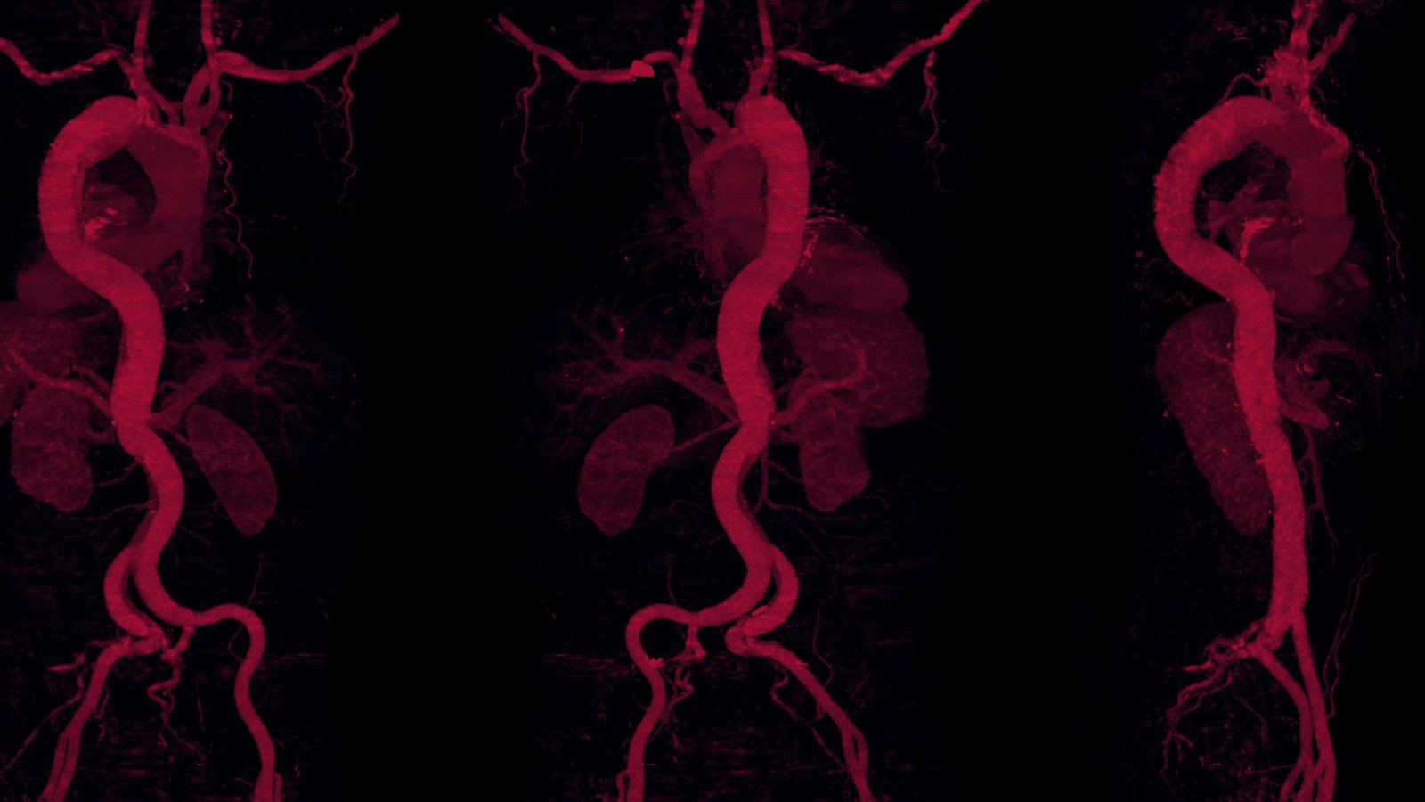
For patients arriving in the ER with an aortic dissection, every minute matters. Delays in diagnosis can allow the weakened aorta to rupture, turning a surgical emergency into a fatal event.
Artificial Intelligence (AI) platforms can rapidly analyze computed tomography angiography (CTA) scans, flag suspected aortic dissections, and alert care teams to prioritize urgent cases, helping reduce delays that can prove fatal.
Aortic dissection, the third-leading noncardiac cause of sudden unexpected death, carries a 1%–2% increase in mortality per hour in the first 48 hours if left untreated.1,2 Because rapid detection and accurate diagnosis are so vital to ensuring patient survival, AI algorithms that recognize the features of an aortic dissection via imaging, such as dissection flaps and aneurysms, can generate real-time alerts that help mobilize surgical teams more quickly, mitigating risks of mortality.
A large-scale validation study that examined more than 1,300 chest and thoraco-abdominal CT angiography images revealed that an AI-powered software platform showed an 80.1% positive predictive value for aortic dissection.3
Another study reviewed an aortic dissection algorithm using the noncontrast-enhanced CT images of 170 patients.4 Researchers found that the algorithm had an “area under the curve of 0.940 for detecting aortic dissection, with an accuracy of 90.0%, sensitivity of 91.8%, and specificity of 88.2%.”4 According to the researchers, the algorithm “showed comparable diagnostic performance to radiologists for detecting aortic dissection.”
While AI-enhanced triage can lead to improved patient outcomes, it is essential to understand the specific pathology of the condition that is being assessed.
A digital subtraction angiography of the abdominal aorta with a stent graft featuring a Cydar image fusion overlay. Red rings identify the renal, visceral, and iliac vessel origins, as well as the proximal aortic landing zone.
Whether identifying a tear algorithmically or interpreting a scan manually, physicians must accurately distinguish between the two types of aortic dissection: Type A begins in the ascending aorta and requires immediate treatment due to life-threatening complications; Type B, which originates in the descending aorta, generally is less immediately life-threatening than Type A, although Type B aortic tears can lead to mortality without medical intervention.
“If we look at how things were done historically, patients entering the ER received a 2D scan for aneurysmal disease, referrals were made, and surgery was performed based on that surgeon’s 3D creation in their mind of a 2D image in terms of where to place stents,” said Allan M. Conway, MD, FACS, a vascular surgeon and associate professor of surgery in the Division of Vascular and Endovascular Surgery at University of California San Francisco Health and MarinHealth.
Traditionally, assessment for aortic dissection has been a manual process that depends on individual expertise and interpretation. Emerging AI tools can help augment and streamline this approach.
“I think where AI has the potential to really change the game is by allowing us to recognize these pathologies much faster and, as a result, increase the efficiency of the referral process,” he added. “In this scenario, when a patient comes into the ER with symptoms, the scan is done, AI recognizes the pathology and alerts the surgical team, helping get operating rooms ready and ultimately decreasing the time from presentation to the operation.”
Dr. Allan Conway and Tina Desai, MD, FACS, in the OR after completing their first case using Cydar Maps technology in 2024.
Digital Twins and Surgical Planning
As AI algorithms grow increasingly proficient at interpreting diagnostic images, researchers are exploring how those data can be used to generate patient-specific virtual models, also known as digital twinning, to explore potential treatment pathways before a procedure begins.
The concept of twinning originated in aerospace engineering, where virtual models are stress-tested under conditions too costly, or impossible, to reproduce physically.
“While digital twinning is a fairly new concept in medicine, it’s well known that engineers will stress jet engines to see how they will respond in real-world scenarios,” explained Dr. Conway. “We’re starting to see this in the surgical world. Ideally, what we want to do is ‘stress’ the patient virtually. So, for aortic dissection repair, we would try different stents and treatment types to see how the virtual patient responds.”
While largely in the experimental phase in medicine, the intention behind twinning is to offer clinicians treatment pathways that are data-driven, adaptive, and personalized. A scientific article published in 2025, “Digital twins for the era of personalized surgery,” describes this approach as follows: “A digital twin in surgery is a dynamic virtual replica of an individual’s physical and physiological state, integrating both bodily systems and healthcare interactions.”5 This technology, which could be used in the preoperative, intraoperative, and postoperative stages, is an evolution of other advanced tools, including 3D modeling and virtual reality simulations.
“For aortic work, I think twinning has the ability to change the way in which we treat patients. For most aortic work, it’s typically an individual surgeon making a plan,” said Dr. Conway. “I think where twinning will help, and this will rely on good data, is I should be able to have a CT scan analyzed by AI software and it should be able to predict how that aneurysm or dissection is going to behave with different treatment models and whether that’s a nonoperative approach versus an X, Y, or Z type of stent, and how that patient is going to respond 1 year, 2 years, and 5 years down the line.”
The surgical team reviews the image fusion overlay before stent graft deployment. The live fluoroscopy image, highlighted by a yellow outline, is paired with red rings identifying critical vascular landmarks to support image-guided aortic surgery.
A January 2026 study examined the accuracy of a thoracic endovascular aortic repair (TEVAR) digital twin for preoperative planning. TEVAR is considered the gold standard for complicated acute Type B aortic dissections depending on the location and acuity of the dissection. Researchers retrospectively analyzed pre- and postoperative CTA scans from patients treated with a specific stent graft. Using a computer simulation of 20 patients to determine how accurately it could predict the placement of the stent graft, researchers found the simulation closely matched what happened in the human patients.6
On average, the difference between the simulation and the actual outcome was only about 4%.6 “The integration of such a validated methodology into routine clinical workflows within a very short simulation time could reduce procedural risks, enhance precision in device deployment, and ultimately improve patient outcomes,” the study’s authors noted.
“I think we’re going to start seeing this approach in the next wave of aortic care,” said Dr. Conway. “In fact, surgeons are already doing it.”
While widespread use of digital twinning, which can be thought of as a virtual dress rehearsal, is still on the horizon, this tool has demonstrated use in surgical specialties such as orthopaedics, neurosurgery, and plastic and reconstructive surgery—specifically for procedures where precision is essential and anatomical structures can vary greatly.
“I think one of the largest applications of AI will be in preoperative decision-making,” added Jason T. Lee, MD, FACS, chief of the Division of Vascular Surgery in the Department of Surgery at Stanford Medicine in Palo Alto, California. Dr. Lee’s focus is on minimally invasive diagnostics, aortic surgery, and complex endovascular aneurysm repair. “Now that hundreds of thousands of implants for TEVAR have been performed worldwide over the last 35 years, we have amassed a bank of outcomes data. Researchers are analyzing imaging findings, preoperative metrics, electronic health records, and other large datasets to determine which patients are the best candidates for these procedures.”
Despite the promising ability of digital twinning and other preoperative assessments to test, predict, and optimize surgical approaches, barriers to universal adoption persist, including high costs, developing standardized validation models, and integrating vast amounts of patient data into these platforms.
“As healthcare systems generate increasingly large and complex datasets, AI offers the computing power needed to analyze information that would otherwise be difficult to manage,” said Dr. Lee. “Most surgeons are not concerned that AI will replace them in the OR. Instead, we see the value it brings by augmenting routine yet important tasks, such as preoperative risk assessment and postoperative follow-up. The more AI can support those efforts, the better care we can provide.”
GPS for Complex Aortic Repairs
Once a treatment strategy has been determined—whether guided by conventional imaging, AI-enhanced analysis, or patient-specific digital simulations—the next challenge is executing that plan in the OR.
Just as AI can help surgeons interpret complex imaging data and identify optimal treatment approaches, this technology also can assist in translating those insights into precise intraoperative action. Specifically, advanced image-guidance platforms can assist surgeons performing complex, minimally invasive vascular surgeries, including aortic dissection repairs.
Using preoperative CT scans, this technology generates precise 3D anatomical models that are superimposed onto live x-ray screens (fluoroscopy) in the OR. This technique, referred to as image fusion technology, has evolved into an important tool used in complex endovascular interventions. Several image fusion platforms are available, including Philips VesselNavigator and Cydar Maps.
According to Dr. Conway, Cydar’s distinguishing feature is its cloud-based structure, which uses AI to automatically map patient-specific aortic anatomies during surgery, including the location of the aneurysm and critical branch vessels, such as the renal, celiac, superior mesenteric, and left subclavian arteries.
“We have been using it for all our aortic cases since 2024,” said Dr. Conway. “When patients ask us about this technology, we assure them that AI is not doing the surgery and that AI is not telling us where to place our stents and where to place our wires. We’re still the decision-makers, and we’re still the ones performing the surgery.”
In addition to its “GPS-like” guidance overlays that enhance surgical precision, image fusion platforms also show measurable gains in efficiency, with some studies showing a reduction in procedure time by as much as 20%.7
“I think knowing where the vessels are where we need to put the x-ray machine in advance really speeds up a lot of the imaging that we have to acquire,” said Dr. Conway, who also noted a reduction in radiation exposure for everyone in the OR (patient and staff) by as much as 50%.7 “As soon as you put your foot on the pedal and start using fluoroscopy, radiation is being delivered. With image fusion technology, there is less radiation time because you know where the vessels are. You also don’t have to inject as much contrast dye to locate the vessels.”
Barriers to Wider Adoption
The precision of fusion mapping relies on high-quality preoperative imaging. “You need good-quality CT scans to be able to produce accurate fusion maps with thin slices that are 1 millimeter or less in thickness in order to capture intricate anatomical details,” explained Dr. Conway.
While high-resolution imaging provides greater anatomical detail, processing and integrating hundreds of CT images requires substantial computational power. The costs associated with processing these data are significant and typically based on an annual subscription model.
“I think one big hurdle with this technology is cost. Convincing hospital administrators to part with the dollars for a yearly recurring fee can be difficult, especially when a lot of these technologies are expensive and there isn’t one technology that fits everybody. We are very grateful for our philanthropic support, which has been instrumental in helping us adopt and use this technology,” said Dr. Conway.
For many institutions, decisions about investing in this technology are based on whether improvements in efficiency, radiation reduction, contrast savings, and workflow justify the recurring expense.
As hospital systems weigh the value of these platforms, surgeons must also consider how to integrate them into practice. “I think there is a risk associated with relying solely on the technology for clinical decision-making, and not using the other tools—fluoroscopy, angiography, anatomical expertise, and procedural experience—that we’ve trained on for years, which could lead to problems. AI technology is a guide,” advised Dr. Conway.
ARISE II Results Highlight Clinical Promise
While advanced imaging and guidance help optimize surgical precision for complicated aortic repair cases, long-term outcomes for these patients will continue to depend on the performance of the endovascular devices themselves.
Although endovascular stent grafts are routinely used to treat descending aortic aneurysms and dissections, there are currently no approved devices for repair in the ascending aorta. The ARISE II trial is a multicenter, National Institutes of Health clinical study, which is following patients for 5 years to evaluate the GORE ascending stent graft. The aim of this pivotal trial is to determine whether this catheter-delivered device can serve as a minimally invasive endovascular alternative to open-heart surgery for patients with ascending aortic disease who face a high risk of surgical complications.
“Stanford Medicine was one of the first sites on the West Coast to treat a patient with this device within the ARISE II trial,” said Dr. Lee. “Surgeons from the Stanford Department of Cardiothoracic Surgery and Stanford’s Division of Vascular & Endovascular Surgery worked in collaboration to test the safety and efficacy of the ascending aortic stent graft for the treatment of conditions such as dissection, pseudoaneurysm, or ulceration of the ascending aorta.”
The 61-year-old patient who was the subject of the Stanford trial had a history of chronic Type B aortic dissection with aneurysmal degeneration. According to a press release issued by Stanford Medicine, the patient received the stent graft without complications and was discharged home after 3 days.8 If the patient had received open-heart surgery, the hospital stay could have lasted as long as 1 week followed by 4–6 weeks of recovery at home.
“We were fortunate that we had a very good outcome for our patient that we submitted for the trial, and we look forward to the next trial to further learn about the appropriate patients and techniques to safely use this ascending stent graft,” Dr. Lee said.
In December 2025, enrollment began in the ARISE III trial, which will assess the same ascending stent graft but in patients requiring emergency repair for Type A dissections. According to Dr. Lee, Stanford Medicine will again be a participant in the trial.
“Endovascular techniques have revolutionized a lot of aortic care during the last 25 years,” said Dr. Lee. “It was natural that eventually we would have devices to potentially use in the ascending aorta, and participating in this trial is just another step in the evolution of us getting closer to the heart itself and expanding our ability to treat these pathologies.”
For Dr. Lee, exploring repair options in the ascending aorta represents the next frontier in a decades-long endovascular revolution. “Developing devices for the ascending aorta is a natural progression as we continue expanding our ability to treat pathology closer to the heart.”
Still, Dr. Lee stressed that open repair remains the gold standard for ascending aortic disease, backed by decades of innovation and excellent long-term outcomes. The promise of an endovascular approach, he said, lies in its potential to complement—not compromise—the durable outcomes achieved through traditional surgery, offering another option for carefully selected patients with challenging anatomy or elevated operative risk.
From AI-powered triage and digital twinning to image fusion guidance and next-generation endovascular devices, emerging technologies are reshaping every stage of aortic care. These innovations offer the potential to accelerate diagnosis, refine procedural planning, enhance intraoperative precision, and expand treatment options for patients once considered too high risk for intervention.
Yet as these tools become more sophisticated, their greatest value will continue to be their ability to augment the surgeon’s clinical judgment. The future of aortic care lies in harnessing data-driven insights while maintaining the human expertise needed to interpret complex anatomy and deliver personalized treatment.
Tony Peregrin is the Managing Editor of Special Projects in the ACS Division of Integrated Communications in Chicago, IL.
References
Huynh-O’Keefe C, Kinkead B, Tsan J, Connolly A, et al. Meeting abstract: Abstracts from the American Heart Association’s 2025 scientific sessions and the American Heart Association’s 2025 Resuscitation Science Symposium. November 3, 2025. Available at: https://www.ahajournals.org/doi/abs/10.1161/circ.152.suppl_3.4367786. Accessed June 15, 2026.
Shubietah A, Elgendy MS, Nazir A, Ahmed A, et al. Aortic dissection mortality in the United States, 1968–2023: Trends, disparities, and deep learning forecasts. Int J Cardiol Cardiovasc Risk Prev. 2025;27:200547.
Salehi S, Schlossman J, Chowdhry S, DeGaetano A, et al. Real-world validation of a deep learning AI-based detection algorithm for suspected aortic dissection. UC Irvine. November 2022. Available at: https://escholarship.org/uc/item/6tm3x7zj. Accessed June 15, 2026.
Hata A, Yanagawa M, Yamagata K, Suzuki Y, et al. Deep learning algorithm for detection of aortic dissection on non-contrast-enhanced CT. Eur Radiol. 2021;31(2):1151-1159.
Mekki Y, Luijten G, Hagert E, Belkhair S. Digital twins for the era of personalized surgery. npj Digit. Med. 2025. Available at: https://www.nature.com/articles/s41746-025-01575-5. Accessed June 15, 2026.
Ramella A, Ded Camp G, Barati S, Heigmen R, et al. Prediction of thoracic endovascular aortic repair intervention: Proof of concept using digital twin technology. Eur J Vasc Endovasc Surg. Published online January 9, 2026. Available at: https://www.ejves.com/article/S1078-5884(26)00013-4/fulltext. Accessed June 15, 2026.
Diagnostic and International Cardiology. Endovascular 3D image guidance added to Philips Zenition Mobile C-Arm. January 18, 2022. Available at: https://www.dicardiology.com/content/endovascular-3d-image-guidance-added-philips-zenition-mobile-c-arm. Accessed June 15, 2026.
Stanford Medicine. Stanford completes first investigational endovascular ascending aorta stent graft in the western US as part of ARISE II national clinical trial. February 1, 2025. Available at: https://med.stanford.edu/ctsurgery/about-the-department/news/2025/first-endovascular-ascending-aorta-stent-graft-in-western-us.html. Accessed June 15, 2026.
HPE announced an expanded collaboration with Oracle to help scale Oracle’s global AI infrastructure by deploying HPE Juniper Networking across Oracle’s AI data centers. The expanded collaboration builds on more than a decade of engineering work between Oracle and Juniper Networks and includes networking support services and financing capabilities. Also Read: AiThority Interview with Gou Rao, co-founder
/PRNewswire/ -- MediaGo, the intelligent advertising platform, has won the Excellence Award in the AdTech category at the 2026 Global Tech Awards. Marking the third consecutive year receiving this honor, the recognition underscores MediaGo's continued innovation in deep learning-powered digital marketing solutions and the tangible business value delivered to advertisers worldwide. The Global Tech
Gene editing has promised a way to treat some diseases, including inherited genetic disorders, blood conditions and cancer, at their genetic roots. But even as tools such as CRISPR, which allows scientists to alter DNA sequences and modify gene function with high precision, have become increasingly powerful, a practical challenge remains: Many genome editors are
We use cookies on our website to give you the most relevant experience by remembering your preferences and repeat visits. By clicking “Accept”, you consent to the use of ALL the cookies.
This website uses cookies to improve your experience while you navigate through the website. Out of these, the cookies that are categorized as necessary are stored on your browser as they are essential for the working of basic functionalities of the website. We also use third-party cookies that help us analyze and understand how you use this website. These cookies will be stored in your browser only with your consent. You also have the option to opt-out of these cookies. But opting out of some of these cookies may affect your browsing experience.
Necessary cookies are absolutely essential for the website to function properly. This category only includes cookies that ensures basic functionalities and security features of the website. These cookies do not store any personal information.
Any cookies that may not be particularly necessary for the website to function and is used specifically to collect user personal data via analytics, ads, other embedded contents are termed as non-necessary cookies. It is mandatory to procure user consent prior to running these cookies on your website.