There is something deep in our nature that is always fascinated by transformations. Artificial intelligence (AI) made it possible for just about anyone with internet access to instantly swap faces of people. And the results are now more realistic than they have ever been. By using the best AI face swap tools, the results are
AI-driven lung imaging: Advancing early diagnosis and monitoring
Standard methods for diagnosing lung diseases often only detect problems once damage has already occurred. Learn how AI-driven lung imaging technologies can provide a way to identify disease earlier, allowing for more targeted interventions
Respiratory diseases remain a leading cause of morbidity and mortality worldwide, (1) yet our ability to detect and monitor lung disease at an early stage remains surprisingly limited.
In many cases, clinically meaningful changes in lung function occur long before they are detectable using standard tools, delaying intervention and contributing to irreversible damage. This is particularly evident in chronic conditions such as cystic fibrosis, asthma, and bronchiectasis, where disease progression is often patchy and evolves silently over time.
Traditional assessments such as spirometry provide only a global measure of lung function, offering little insight into how disease is distributed within the lungs. While spirometry remains a cornerstone of respiratory care, it lacks sensitivity to early or regional disease and can remain within normal ranges despite significant underlying pathology. (2)
Similarly, computed tomography (CT) provides high-resolution structural information, but does not directly measure how air moves through the lung. As a result, clinicians are often forced to infer function from structure, or detect disease only once structural damage has become established and irreversible.
This gap between structure and function represents one of the most significant challenges in respiratory medicine, (3) and has led to the development of functional lung imaging methods that aim to describe how the lung is working, rather than what it looks like. (4)
AI is transforming lung imaging
Artificial intelligence (AI), particularly machine learning, is now enabling a fundamental shift in how lung imaging data can be analysed and interpreted. Rather than relying solely on visual inspection, AI systems can be trained to identify complex spatial and temporal patterns across large datasets, detecting subtle and early changes that may not be apparent to the human eye. (5)
In the context of lung imaging, AI enables a transition from qualitative interpretation to quantitative, high- resolution assessment of regional lung function. (6)
Importantly, AI allows the integration of both spatial and temporal dynamics, capturing not just what the lung looks like but also how it behaves. This opens the door to tracking disease progression at a much earlier stage, identifying regional dysfunction, and monitoring response to therapy with far greater precision.
These approaches are not intended to replace clinicians but to augment clinical decision-making by providing objective, reproducible, and clinically meaningful metrics.
From structure to function: The role of X-ray velocimetry
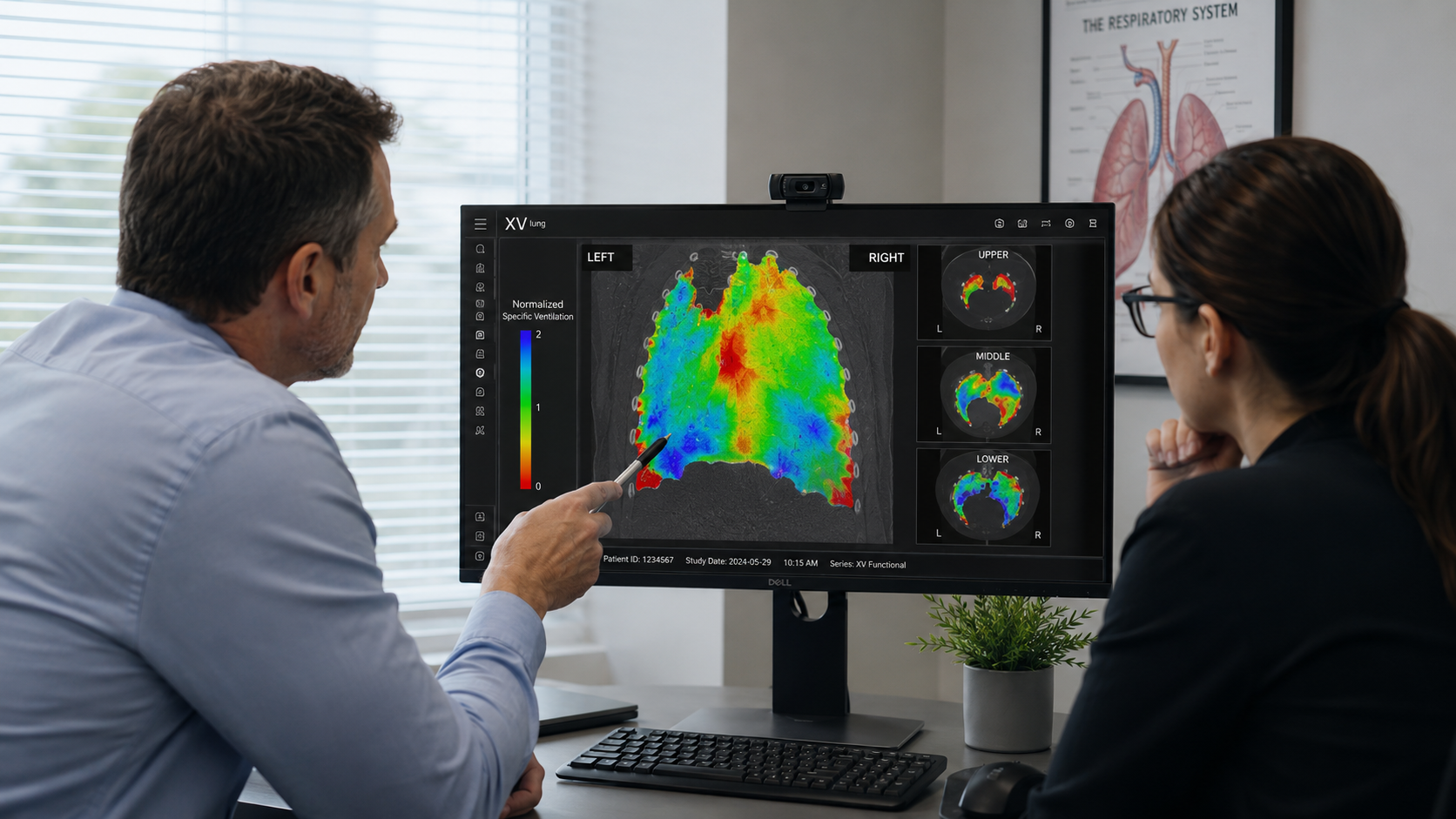
Together, 4DMedical Ltd, Adelaide University, and the University of Melbourne are developing and applying X-ray velocimetry (XV), a novel functional lung imaging technology that directly measures regional ventilation. (7)
XV uses fluoroscopic imaging acquired from multiple angles to track lung motion throughout the breathing cycle.(8) Advanced computational modelling is then used to reconstruct airflow patterns across the lung, generating detailed maps of regional ventilation.(9)
By combining XV with AI-driven analysis, we can move beyond traditional global metrics and directly quantify how different regions of the lung contribute to overall function. Machine learning approaches are being used to characterise airflow patterns, identify disease-specific signatures, and detect subtle changes over time. This is particularly valuable in conditions such as cystic fibrosis, primary ciliary dyskinesia, and asthma, where early disease is highly heterogeneous and may be missed by conventional tests.
A key advantage of this approach is its potential to detect disease earlier and guide more targeted intervention. Rather than waiting for global lung function to decline, clinicians can identify emerging regional abnormalities and intervene before irreversible damage occurs. (10) In addition, these techniques offer a powerful tool for evaluating therapeutic response, supporting both clinical care and the development of new treatments.
Translating innovation into clinical practice
Our work is supported by funding from Australia’s Economic Accelerator (IV240100090), which is enabling the translation of XV from a research tool into clinical practice. This includes the development of dedicated XV scanner hardware, streamlined imaging protocols, automated analysis pipelines, and integration into existing clinical workflows. Importantly, this program also incorporates engagement with patients, clinicians, and healthcare systems to ensure that the technology is not only effective but also practical, acceptable, and scalable.
Towards precision respiratory medicine
Looking ahead, AI-driven functional lung imaging has the potential to underpin a new model of precision respiratory medicine. By combining detailed regional information with longitudinal data and clinical context, these approaches can support more personalised treatment strategies and improved disease monitoring. As datasets grow and models become more sophisticated, there is also potential to integrate imaging with other modalities, including genomics and digital health technologies.
Ultimately, the goal is to shift respiratory care from reactive – where disease is detected after damage has occurred – to proactive identification and management at its earliest stages. AI-driven imaging technologies such as XV represent an important step towards this future, offering the potential to improve outcomes for patients with respiratory disease both in Australia and globally.
Contributing authors
Martin Donnelley, Tom Drummond, Krista Ehinger, Dhani Dharmaprani, Ronan Smith, Nicole Reyne, and Shreshail Bhatta.
References
- Momtazmanesh, S. et al. Global burden of chronic respiratory diseases and risk factors, 1990–2019: an update from the Global Burden of Disease Study 2019. eClinicalMedicine 59 (2023). https://doi.org/10.1016/j.eclinm.2023.101936
- Pellegrino, R. et al. Interpretative strategies for lung function tests. European Respiratory Journal 26, 948-968 (2005). https://doi.org/10.1183/09031936.05.00035205
- Kirby, M. & Smith, B. M. Quantitative CT Scan Imaging of the Airways for Diagnosis and Management of Lung Disease. Chest 164, 1150-1158 (2023). https://doi.org/10.1016/j.chest.2023.02.044
- Bayat, S., Wild, J. & Winkler, T. Lung functional imaging. Breathe 19 (2023). https://doi.org/10.1183/20734735.0272-2022
- Topol, E. J. High-performance medicine: the convergence of human and artificial intelligence. Nature Medicine 25, 44-56 (2019). https://doi.org/10.1038/s41591-018-0300-7
- Ardila, D. et al. End-to-end lung cancer screening with three-dimensional deep learning on low-dose chest computed tomography. Nature Medicine 25, 954-961 (2019). https://doi.org/10.1038/s41591-019-0447-x
- Vliegenthart, R., Fouras, A., Jacobs, C. & Papanikolaou, N. Innovations in thoracic imaging: CT, radiomics, AI and x-ray velocimetry. Respirology 27, 818-833 (2022). https://doi.org/10.1111/resp.14344
- Bruorton, M. et al. Pilot study of paediatric regional lung function assessment via X-ray velocimetry (XV) imaging in children with normal lungs and in children with cystic fibrosis. BMJ Open 14 (2024). https://doi.org/10.1136/bmjopen-2023-080034
- Smith, R. et al. Mapping lung cancer ventilation dynamics using functional imaging and lung mechanics. Disease Models & Mechanisms 18 (2025). https://doi.org/10.1242/dmm.052559
- Vij, N. Prognosis-Based Early Intervention Strategies to Resolve Exacerbation and Progressive Lung Function Decline in Cystic Fibrosis. Journal of Personalized Medicine 11 (2021). https://doi.org/10.3390/jpm11020096

Related Posts