Key Takeaways Hyperspectral imaging captures extensive spectral data, enabling precise material and tissue differentiation beyond conventional imaging capabilities. Applications span counterfeit detection, environmental monitoring, agriculture, food quality, and medical diagnostics, with significant accuracy improvements. AI and deep learning enhance HSI's analytical potential, addressing challenges like high costs and complex data analysis. The convergence of AI
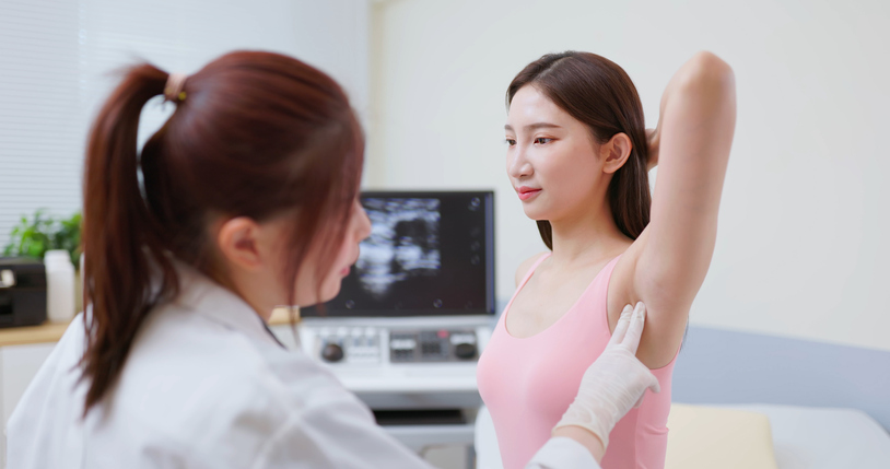
Deep Learning Flags Breast Cancer Risk Between Triennial Screenings

A deep learning algorithm can spot women who might benefit from extra breast cancer imaging in between the triennial screenings offered by the UK National Health Service.
The Mirai tool was able to help determine who should undergo supplemental imaging or have shorter screening intervals to aid the detection of interval cancers (ICs), which occur after a negative screening but before the next screening round.
“Our results suggest that further workup of mammograms within the highest 20% of scores could yield 42.4% of ICs, meaning that Mirai could be used instead of or in addition to breast density to identify women who may benefit from supplemental imaging or a shortened screening interval,” reported Fiona Gilbert, PhD, from the University of Cambridge, and co-workers.
The findings are published in Radiology.
Deep learning algorithms for personalized breast cancer screening have performed better than traditional methods in earlier retrospective studies, but there remains a lack of information on triennial screening assessments.
ICs consist of either missed cancers—false negatives—or new ones that have appeared in the interim before the next screening, and they have a worse prognosis than cancers detected during screening.
Gilbert and team used the Mirai algorithm, developed at Massachusetts General Hospital, to predict interval cancers from mammograms in the UK breast screening program, which invites women aged 50 to 70 years for triennial mammography.
Mirai was retrospectively applied to 134,217 digital mammograms obtained at two sites in the U.K. using two different mammography systems over a three-year period.
Mirai provided a three-year risk score for IC for each mammogram.
Negative digital screening mammograms—where no cancer was found—were analyzed by Mirai in terms of characteristics such as tumor features and breast density to produce a generalized risk score for developing an interval breast cancer.
For each of the 524 interval cancers that occurred, the screening mammogram beforehand was retrospectively reviewed to determine whether it was a “true” case of IC, in which the previous screening mammogram was normal or benign, or “false” ICs in which the screening mammogram was suspicious or uncertain.
Results showed that the three-year risk scores retrospectively predicted 3.6% of ICs (19 of 524) among women assigned the highest one percent of scores, 14.5% (76 of 524) of those for women in the highest five percent of scores, 26.1% (1237 of 524) for those in the highest 10% of scores, and 42.4% (222 of 524) of ICs for those in the top 20% of scores.
Identification of ICs was equivalent to a corresponding extra cancer detection rate of 0.1, 0.6, 1.0, and 1.7 per 1000.
There was no evidence of differences in performance between the 1-, 2-, and 3-year predictions or across age categories or breast densities.
However, Mirai was better at predicting ICs within a year of the screening examination compared with later periods. Although it was less effective in women with extremely dense breast tissue, the algorithm was better than conventional risk prediction tools.
“These results using the deep learning model Mirai to identify women at increased risk are encouraging and offer a potentially important step toward tailored precision breast cancer screening for women,” wrote Liane Philpotts, PhD, from Yale School of Medicine, in an editorial accompanying the study.
“Nevertheless, even with the highest threshold, more than 50% of ICs would not be predicted.
“This humbling fact underscores the difficulty—for both humans and machines—of predicting and detecting breast cancers with mammography.”
Related Posts