Abstract The rapid development of AI-based products and their underlying models has led to constant innovation in deep learning frameworks. Google has been pioneering machine learning usage across dozens of products. Maintaining the multitude of model source codes in different ML frameworks and versions is a significant challenge. So far the maintenance and migration work
Wayne State researchers use AI to predict glaucoma progression and visual field loss

A research duo at Wayne State University is working to develop an artificial intelligence-based framework to better predict visual-field outcomes in glaucoma patients.
The project is supported by a one-year, $38,914 Kresge Eye Institute Translation Research Innovation Grant, an internal pilot grant program sponsored jointly by KEI and the School of Medicine’s Department of Ophthalmology, Visual and Anatomical Sciences.
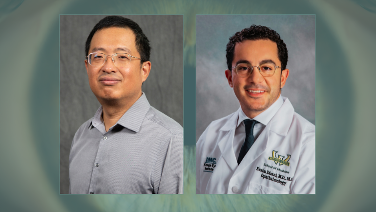
“Predicting Visual Field Progression from Structural and Functional Measurements Toward Multimodal Personalized Glaucoma Modeling,” co-led by Ophthalmology resident Karim Dirani, M.D. ’22, MPH, and College of Engineering Professor of Computer Science Ming Dong, Ph.D., will integrate visual-field and optical coherence tomography measurements to estimate uncertainty in those predictions, with the longer-term goal of developing personalized, multimodal models of glaucoma progression.
Glaucoma is a leading cause of irreversible blindness, making predicting future progression particularly important, Dr. Dirani said. The clinical challenge is that glaucoma doesn’t progress at the same rate in every patient.
“Glaucoma causes gradual and irreversible damage to the optic nerve, but one of the biggest challenges we face is determining which patients are likely to lose vision quickly and which are likely to remain stable,” Dr. Dirani said. “In glaucoma care, we routinely collect several types of information, particularly visual field testing, which measures how well a patient can see across different areas of their vision, and optical coherence tomography, or OCT, which measures the structural health of the optic nerve and retina.”
Current tests only provide pieces of the picture.
“OCT tells us about structural damage to the retinal nerve fiber layer and optic nerve, while visual-field testing tells us how that damage is affecting a patient’s functional vision. The relationship between those two is complicated, differs across areas of the eye and changes depending on the stage of the disease,” he said. “If we can more accurately integrate that information and identify patients who are likely to progress rapidly before substantial vision loss occurs, it could eventually help clinicians personalize monitoring and treatment intensity. It may also allow us to move glaucoma care away from simply documenting what has already happened toward anticipating what is likely to happen next.”
The AI model is being designed to provide a range of possible outcomes and indicate how confident it is in the prediction. While it is focused on prediction, not treatment recommendations, the longer-term goal is to incorporate additional information such as intraocular pressure and treatment history so that future models could potentially simulate how a patient’s disease trajectory might differ under different clinical scenarios. For example, if two patients appear relatively similar during today’s visit but the model identifies one as having a substantially greater probability of rapid future visual-field deterioration, that information could potentially influence how closely the patient is monitored or how aggressively treatment is considered.
“The pilot work is specifically intended to generate preliminary data and establish the methodological foundation for a subsequent NIH R01 application,” said Associate Chair and Professor of Ophthalmology, Visual and Anatomical Sciences Elizabeth Berger, Ph.D. “The KEI-TRIG grant is designed to support innovative, collaborative research that can generate preliminary data for future competitive extramural grant applications.”
The project, Dr. Berger added, leverages a large longitudinal glaucoma dataset developed at KEI of approximately 25 years of clinical data encompassing 43,000 patients, 150,000 visual fields and 160,000 OCT studies, and combines KEI’s clinical expertise with Dr. Dong’s expertise in advanced AI and machine-learning approaches.
“I am extremely grateful to Kresge Eye Institute and the Department of Ophthalmology, Visual and Anatomical Sciences for supporting this work,” Dr. Dirani said. “What is especially exciting about a pilot grant like the KEI-TRIG is that it gives us the opportunity to take an idea that has developed from years of clinical data collection and preliminary research and begin building something that could ultimately have a meaningful impact on patient care.”
Dr. Dirani is the clinical and scientific lead for the project, developing the study design, curation and clinical interpretation of the Kresge glaucoma dataset, defining clinically meaningful outcomes, overseeing model development and validation, and ultimately translating the results back into questions relevant to the care of patients with glaucoma. He has worked extensively with the dataset on earlier studies of glaucoma progression and AI-based visual field prediction, which provided much of the preliminary foundation for this project.
Considered an expert in generative AI, deep learning, graph-based modeling, computer vision and medical image analysis, Dr. Dong will lead and advise the development of the advanced computational architecture, particularly the graph-based and diffusion-modeling components that allow the team to integrate OCT and visual-field information, and model multiple possible future disease trajectories rather than producing only a single prediction.

Related Posts